Back Of Skull Anatomy / Very Detailed And Scientifically Correct Human Skull Back View Stock Photo Picture And Royalty Free Image Image 11713062 : The bbc is not responsible for the content of external websites.
Back Of Skull Anatomy / Very Detailed And Scientifically Correct Human Skull Back View Stock Photo Picture And Royalty Free Image Image 11713062 : The bbc is not responsible for the content of external websites.. Skull, skeletal framework of the head of vertebrates, composed of bones or cartilage, which form a unit that protects the brain and some sense organs. Norma basalis ( anterior part , middle part and posterior part ). The base of the skull (or skull base) forms the floor of the cranial cavity and separates the brain from the structures of the neck and face. Learn vocabulary, terms and more with flashcards, games and other study tools. The skull bones can be classified into two groups:
Cranium) is the skeleton of the head composed of 22 separate bones joined together primarily by sutures. So, the human skull consists of 23 bones. Overview, anterior skull base, middle skull base march 18, 2017. Please feel free to download and print. The cranium and the mandible.
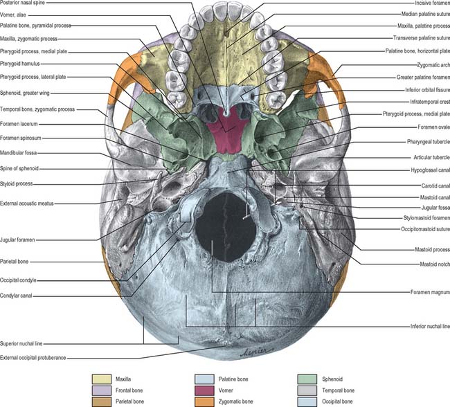
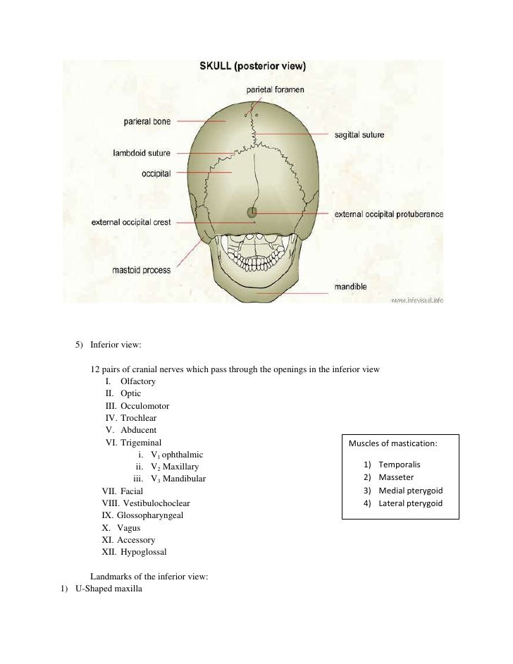
External Skull Clinical Gate from clinicalgate.com The bone is pierced by a large oval hole(the foramen magnum) through which runs the spinal cord. William is a final year medical student in australia who has taught anatomy to tertiary science and. The cranium and the mandible. The skull bones can be classified into two groups: Skeleton anatomy easy review for practical exam bones and structures. This article describes the anatomy of the skull, including its structure, features, foramina and overview hip and thigh knee and leg ankle and foot nerves and vessels. So, the human skull consists of 23 bones. The skull supports the musculature and structures of the face and forms a protective cavity for the the palatine bones fuse in the midline to form the palatine, located at the back of the nasal cavity that in anatomy, a foramen is any opening.
But it's not all bones!
In order to be light, the skull is made up by flat and irregular bones, and has hollow spaces called the sinuses. Cranium) is the skeleton of the head composed of 22 separate bones joined together primarily by sutures. Skull bones aren't fused together at birth. It is comprised of many bones, formed by intramembranous ossification, which are joined together by sutures (fibrous joints). The base of the skull (or skull base) forms the floor of the cranial cavity and separates the brain from the structures of the neck and face. This anatomic region is complex and poses surgical challenges for otolaryngologists and neurosurgeons alike. Foramina inside the body of humans and other animals. Please feel free to download and print. A thorough description is beyond the. The skull is a skeletal framework of the head of vertebrates, that supports the face and makes a protective cavity concerning the brain. The occipital bone forms the back of the skull and the base of the cranium. The two fontanels located on the sides of the skull are mirror. It supports and protects the face and the brain.
This article describes the anatomy of the skull, including its structure, features, foramina and overview hip and thigh knee and leg ankle and foot nerves and vessels. Skull reshaping is done on any of the structures that lie above the face. The skull bones can be classified into two groups: The skull is a bony structure that supports the face and forms a protective cavity for the brain. The base of the skull (or skull base) forms the floor of the cranial cavity and separates the brain from the structures of the neck and face.
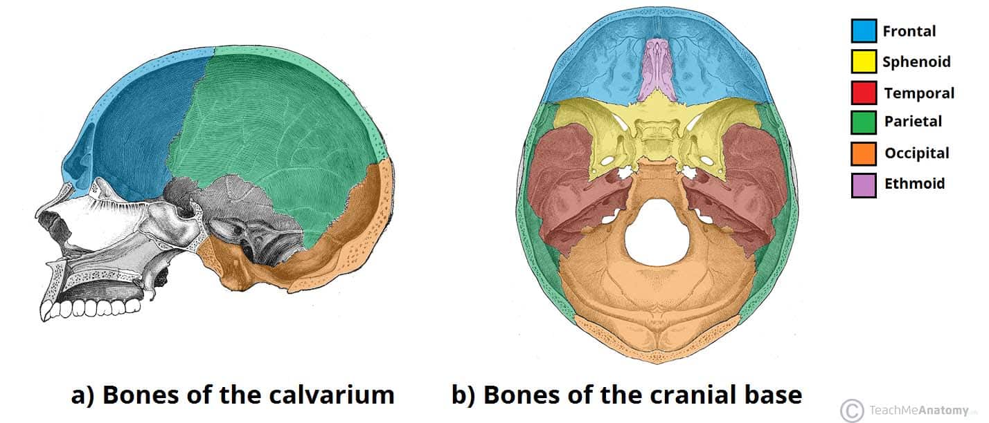
Bones Of The Skull Structure Fractures Teachmeanatomy from teachmeanatomy.info The frontal, parietal, temporal and occipital bones are joined at the cranial sutures. The skull base is the inferior portion of the neurocranium. Please feel free to download and print. It is comprised of many bones, formed by intramembranous ossification, which are joined together by sutures (fibrous joints). It was then cleaned, adapted and polypainted this model is part of a comparison with the skull of a human. The two fontanels located on the sides of the skull are mirror. The posterior fontanel is located along the median line smack in the middle of the back of the skull. Excluding ear ossicles, it is made of 22 bones.
The bbc is not responsible for the content of external websites.
The frontal, parietal, temporal and occipital bones are joined at the cranial sutures. The cranium and the mandible. The axial & appendicular skeleton. Skeleton anatomy easy review for practical exam bones and structures. A cartilaginous mould begins to grow this is why raising your eyebrows can create the appearance that the back of the head is moving. The skull has a single occipital condyle.7 the skull consists of five major bones: Excluding ear ossicles, it is made of 22 bones. From an anatomical perspective, the skull is divided into two parts: The skull begins to form prior to week 12 of embryogenesis. The skull bones can be classified into two groups: It offers protection to the brain, eye balls, inner ears, and nasal passages. Learn about the anatomy of the skull bones and sutures as seen on ct images of the brain. It supports and protects the face and the brain.
Skull, skeletal framework of the head of vertebrates, composed of bones or cartilage, which form a unit that protects the brain and some sense organs. The axial & appendicular skeleton. The skull is the bony skeleton of the head. Anatomy next provides anatomy learning tools for students and teachers. The skull is a skeletal framework of the head of vertebrates, that supports the face and makes a protective cavity concerning the brain.
Skull Notes from image.slidesharecdn.com Cranial cavity , cranial sutures. So, the human skull consists of 23 bones. Anatomy of the skull and bones of cranium on medical illustrations. The two fontanels located on the sides of the skull are mirror. The axial & appendicular skeleton. Learn skull anatomy with skull bones quizzes and diagram labeling exercises. The skull or known as the cranium in the medical world is a bone structure of the head. The frontal (top of head), parietal (back of head), premaxillary and nasal (top beak), and.
It offers protection to the brain, eye balls, inner ears, and nasal passages.
Looking at it from the inside it can be subdivided into. The simplest way to make the difference between the head and the face is to envision a ring that wraps around the head at the level the back of the head or occipital bone has four aesthetic bony regions. Anatomy of the skull and bones of cranium on medical illustrations. The skull begins to form prior to week 12 of embryogenesis. These joints fuse together in adulthood. Overview, anterior skull base, middle skull base march 18, 2017. In order to be light, the skull is made up by flat and irregular bones, and has hollow spaces called the sinuses. The bone is pierced by a large oval hole(the foramen magnum) through which runs the spinal cord. The skull has a single occipital condyle.7 the skull consists of five major bones: The skull base is the inferior portion of the neurocranium. Please feel free to download and print. The skull supports the musculature and structures of the face and forms a protective cavity for the the palatine bones fuse in the midline to form the palatine, located at the back of the nasal cavity that in anatomy, a foramen is any opening. The frontal, parietal, temporal and occipital bones are joined at the cranial sutures.


0 Komentar